Nail IABP on CCRN: 3 High Yield Facts, Waveforms, Weaning
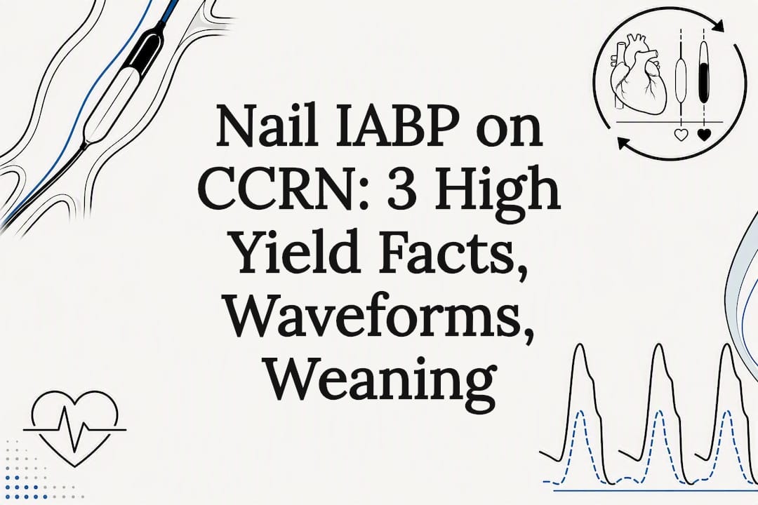
An intra-aortic balloon pump reduces afterload during systole and boosts coronary perfusion during diastole by inflating and deflating in counterpulsation with the cardiac cycle. For the CCRN exam, memorize three things cold: the balloon tip sits about 2 cm distal to the left subclavian artery at the level of the carina, inflation begins at the dicrotic notch and deflation happens just before systole, and limb ischemia is the complication you’ll be tested on most.
TL;DR:
- The intra-aortic balloon pump tip should sit about 2 cm below the left subclavian artery at the level of the carina, confirmed by chest x-ray.
- Inflation occurs at the dicrotic notch during diastole to augment coronary perfusion, while deflation just before systole decreases afterload.
- Limb ischemia is the most tested complication and requires hourly pulse checks with prompt vascular intervention if perfusion diminishes.
- Contraindications include aortic dissection and severe aortic regurgitation; insertion requires careful positioning and waveform troubleshooting.
- Hemodynamic targets include maintaining MAP above 65 mmHg, improving urine output, and observing a downward lactate trend during support.
Table of Contents
- What Is IABP on the CCRN Exam? Physiology and Hemodynamic Effects
- When Is an IABP Indicated, and When Is It Contraindicated?
- How Is an IABP Inserted, and What’s the Correct Position?
- How Do You Read IABP Waveforms and Fix a Bad Trace?
- What Should You Monitor While a Patient Is on IABP?
- What Are the Major IABP Complications, and How Do You Respond?
- When Can You Wean and Remove an IABP?
- High-Yield IABP Facts for CCRN Exam Recall
- A Nurse-to-Nurse Take on IABP Care
- Master IABP and the Rest of Cardiovascular With Zero Deficit™
- Where This Article’s Facts Come From
- Sources
- FAQ
What Is IABP on the CCRN Exam? Physiology and Hemodynamic Effects
The intra-aortic balloon pump works through a principle called counterpulsation. The balloon inflates during diastole and deflates right before systole, and that timing is the entire mechanism you need to understand for both patient care and exam questions.
During diastole, balloon inflation displaces blood volume in the aorta and raises diastolic pressure. That pressure spike, called diastolic augmentation, pushes blood backward into the coronary arteries, which fill almost exclusively during diastole in the first place. The StatPearls review confirms this is the core benefit: augmented coronary perfusion in a heart that’s struggling to perfuse itself.
Deflation is where the second benefit kicks in. Just before the ventricle contracts, the balloon deflates rapidly and creates a vacuum effect in the aorta. That drop in pressure means the left ventricle has to push against less resistance, so afterload falls and myocardial oxygen demand drops with it. You’re essentially giving a sick heart a lighter workload and a better fuel supply at the same time.
At the bedside, you’ll track these effects through a specific cluster of numbers:
- Mean arterial pressure (MAP): should trend upward, ideally sustained above 65 mmHg
- Diastolic augmentation on the arterial waveform: the augmented peak should exceed the unassisted systolic peak
- Urine output: improving output signals better renal perfusion and forward flow
- Lactate: a downward trend suggests improving tissue oxygenation and reduced anaerobic metabolism
Keep in mind the StatPearls authors note IABP’s hemodynamic effect is modest compared with devices like Impella or ECMO. It remains widely used anyway because it’s simpler to insert, manage, and wean, which is exactly why the CCRN exam favors it as a core mechanical circulatory support concept over more complex devices covered in mechanical circulatory support questions.
When Is an IABP Indicated, and When Is It Contraindicated?
The CCRN exam tests indications and contraindications as paired concepts, so you need both sides memorized together, not in isolation.
Accepted indications include:
- Cardiogenic shock, particularly as a bridge to a more definitive therapy or recovery
- Mechanical complications after myocardial infarction, such as acute mitral regurgitation or ventricular septal rupture
- High-risk percutaneous coronary intervention, where hemodynamic support during the procedure improves safety
- Postcardiotomy support when a patient can’t wean off bypass
Absolute contraindications you must recognize instantly:
- Aortic dissection
- Severe aortic regurgitation, since the balloon would worsen regurgitant flow during diastole
Relative contraindications include significant peripheral arterial disease, severe aortoiliac occlusive disease, and bleeding disorders that raise the risk of vascular complications at the insertion site.
Guideline framing matters here for exam nuance. The StatPearls guideline discussion notes that ACC/AHA STEMI guidance has historically given IABP a IIa recommendation, meaning it’s reasonable to consider in select cardiogenic shock patients as a temporary stabilizing measure, while European guidance has been more reserved. The CCRN exam won’t ask you to argue for or against IABP use. It will ask you to recognize the scenario where it’s indicated and the scenario where it’s dangerous.
How Is an IABP Inserted, and What’s the Correct Position?
Three components make up the system you’ll manage at the bedside: the balloon catheter itself, a central lumen that allows arterial pressure monitoring and blood gas sampling, and the external console, which controls inflation timing based on a trigger signal, usually the ECG R-wave or the arterial pressure waveform.
Insertion happens percutaneously, almost always through the femoral artery, using a Seldinger technique similar to other arterial line placements. The bedside nurse’s job during and after insertion centers on maintaining strict asepsis at the insertion site and watching for early signs of vascular compromise in the affected limb the moment the sheath goes in.
Confirming correct position is where CCRN questions get specific. Follow these steps after insertion:
- Obtain a portable chest x-ray immediately after placement
- Confirm the balloon tip sits approximately 2 cm distal to the left subclavian artery, at the level of the carina
- Document the confirmed position and communicate it to the team before relying on the waveform for titration
- Reassess position after any patient repositioning, since migration can occur
The IBCC page on IABP treats this radiographic landmark as a mandatory bedside checkpoint, not a one-time formality. A tip that migrates too high risks occluding the left subclavian artery, and one that sits too low reduces effective augmentation and raises the risk of renal or mesenteric ischemia.
Pro Tip: Say the landmark out loud when you chart it: “tip at carina, 2 cm below left subclavian.” Turning the number into a verbal habit at the bedside is exactly what makes it stick for exam recall too.
How Do You Read IABP Waveforms and Fix a Bad Trace?
An effective IABP tracing has a signature look: the diastolic augmentation peak rises higher than the patient’s own unassisted systolic peak, and the assisted end-diastolic pressure drops lower than the unassisted end-diastolic pressure. If you don’t see both of those relationships on the strip, something is off, and the exam expects you to know exactly what to check.

Inflation should occur at the dicrotic notch, which marks aortic valve closure and the start of diastole. Deflation should occur immediately before the next systole, so the ventricle contracts against a fully deflated balloon and minimal resistance. Arrhythmias complicate this constantly, because irregular R waves throw off ECG-triggered timing, which is why many consoles allow switching between ECG and pressure triggers depending on the rhythm.
Work through timing errors in this order:
- Early inflation shows up as augmentation starting before the dicrotic notch, closing the aortic valve prematurely and raising afterload rather than lowering it
- Late inflation shows suboptimal augmentation with a widened dicrotic notch, meaning you’re losing coronary perfusion benefit
- Early deflation produces a sharp drop right after augmentation, followed by a rise before systole, and it reduces the afterload-reduction benefit
- Late deflation raises afterload because the balloon is still inflated when the ventricle tries to eject
Before touching timing settings, confirm the ECG signal is clean and the trigger mode fits the rhythm, according to IBCC’s troubleshooting guidance. A patient in frequent atrial fibrillation may need a pressure trigger instead of ECG, because irregular R waves make ECG triggering unreliable. Once trigger and rhythm are sorted, work through balloon integrity, catheter position, and console alarms in that order before assuming the pump itself has failed.
What Should You Monitor While a Patient Is on IABP?
Continuous arterial pressure monitoring is non-negotiable with IABP, and most units run it through the balloon’s own central lumen or a separate arterial line, giving you a real-time view of both augmented and unassisted pressures on the same tracing.
Effective support shows up clinically, not just on the monitor. Watch for:
- MAP sustained above target, typically 65 mmHg or higher, without escalating vasopressor doses
- Urine output trending upward as renal perfusion improves
- Lactate clearing over serial measurements, reflecting better tissue oxygenation
- Ability to wean vasopressor or inotrope infusions while hemodynamics hold steady
Anticoagulation practice varies by institution and by bleeding risk, but the underlying rationale is consistent: the balloon and catheter surface create a risk of thrombus formation, so many protocols use low-dose heparin or another anticoagulant, balanced constantly against the patient’s bleeding risk from vascular access, surgical sites, or coagulopathy. The CCRN exam expects you to recognize this as a risk-benefit balance rather than a fixed rule, since practice differs across centers.
What Are the Major IABP Complications, and How Do You Respond?
Limb ischemia is the complication you’ll see tested most often, and it’s also the one where fast recognition genuinely changes outcomes.
- Check pulses and perfusion every hour in the limb distal to the insertion site, comparing color, warmth, and capillary refill against the contralateral limb
- Use Doppler immediately if pulses are diminished or absent, and escalate to vascular surgery without delay if ischemia is confirmed, since early intervention improves limb-salvage outcomes according to StatPearls’ complication review
- Watch the insertion site continuously for bleeding and infection, checking dressings, tracking white blood cell trends, and monitoring temperature curves
- Recognize rare but catastrophic events, including balloon rupture, which presents as blood in the balloon tubing, and aortic injury, which can present as sudden hypotension or back pain; both require immediate console shutdown and emergent physician notification
A structured review of mechanical circulatory support devices situates these vascular and bleeding complications as a known tradeoff across IABP and more advanced support devices alike, which is worth knowing when the exam asks you to compare risk profiles.
When Can You Wean and Remove an IABP?
Weaning decisions rest on a checklist, and the CCRN exam tests whether you know the checklist, not just the final removal step.
- Confirm hemodynamic stability off escalating support: stable MAP, adequate perfusion, and a downward lactate trend with minimal or no vasopressor requirement
- Reduce the augmentation ratio stepwise, typically moving from 1:1 to 1:2 or 1:3, watching for hemodynamic deterioration at each step before advancing
- Trial the patient at a lower ratio for a defined period, monitoring MAP, heart rhythm, and clinical signs of low output before proceeding to removal
- Monitor closely after removal, with frequent vascular checks at the insertion site, ongoing bleeding surveillance, and clear documentation of pulses, sensation, and site appearance at each check
A patient who decompensates at any ratio reduction goes back to the prior setting, and that’s a normal, expected part of the process rather than a failure.
High-Yield IABP Facts for CCRN Exam Recall
Three numbers and relationships carry more exam weight than any deep dive into trial data: the tip position at the carina, the dicrotic notch timing for inflation, and the absolute contraindications list. Build your recall around those, not around memorizing outcome statistics from individual studies.
- Tip position: approximately 2 cm distal to the left subclavian artery, at the carina
- Inflation timing: at the dicrotic notch, marking aortic valve closure
- Deflation timing: immediately before systole, minimizing afterload at ejection
- Absolute contraindication: aortic dissection and severe aortic regurgitation
- Most tested complication: limb ischemia, requiring hourly pulse checks
Zero Deficit™'s cardiovascular study guide organizes IABP alongside cardiogenic shock, ACS, and hemodynamics so you’re studying it in the same clinical cluster the exam draws from. Once the physiology is solid, spaced repetition through the CCRN practice question bank is what moves these facts from “recognized” to “automatic” under exam pressure, with detailed rationales explaining why each wrong answer is wrong.
A Nurse-to-Nurse Take on IABP Care
Make position confirmation, waveform checks, and pulse checks part of your routine, not an afterthought triggered by an alarm. A simple mnemonic helps: “notch in, systole out” for timing, and “carina, 2, clavicular” for position. Bedside habits like these are what turn textbook physiology into instinct when a real patient’s pressure is dropping.
— Zero
Master IABP and the Rest of Cardiovascular With Zero Deficit™
You’ve just covered more IABP detail than most review books pack into a single chapter, and that’s exactly the density Zero Deficit™ builds its cardiovascular content around. Instead of a generic overview, you get topic-specific practice questions with detailed rationales explaining not just the right answer but why each distractor is wrong, written by ICU nurses who know where CCRN candidates actually get tripped up on device physiology.
Start with the cardiovascular study guide to see how IABP connects to cardiogenic shock and ACS, then move into the 695-question practice bank to test your recall on timing, positioning, and complications under real exam conditions. Full access is available through subscription options on Zero Deficit’s subscription page, including monthly and yearly plans, as well as a Lifetime Mastery option if you’d rather pay once and keep the material for every recertification cycle ahead. Pick a plan, open your first practice quiz, and start closing the gaps this article just showed you.
Where This Article’s Facts Come From
For deeper reading beyond this guide, the StatPearls IABP review offers a comprehensive clinical overview, the Internet Book of Critical Care covers waveform troubleshooting in practical bedside detail, and the AACN CCRN certification page explains exam eligibility and test plan structure. The LitFL IABP overview is a concise supplementary read for indications and physiology.
Sources
- Intra-Aortic Balloon Pump - StatPearls - NCBI Bookshelf
- Intra-aortic balloon pump (IABP) — Internet Book of Critical Care (IBCC)
- Get Your CCRN Certification - AACN
FAQ
What Does IABP Stand For?
IABP stands for intra-aortic balloon pump, a mechanical circulatory support device that inflates and deflates in counterpulsation with the cardiac cycle. It augments coronary perfusion during diastole and reduces afterload during systole, as described in the StatPearls review.
Is It Safe to Perform CPR With an IABP In Place?
Yes, CPR can be performed with an IABP in place, though most consoles have a CPR mode that adjusts or pauses timing to accommodate chest compressions. Follow your unit’s protocol and the console manufacturer’s instructions, since compressions can otherwise interfere with trigger detection and balloon timing.
Does an IABP Require Anticoagulation?
Anticoagulation practice varies by institution, and there’s no single fixed rule tested on the exam. Many protocols use low-dose heparin to reduce thrombus risk on the catheter surface, balanced against each patient’s individual bleeding risk from vascular access or other sites.
How Do You Interpret an IABP Waveform?
An effective tracing shows a diastolic augmentation peak higher than the patient’s unassisted systolic peak, with a lower assisted end-diastolic pressure than the unassisted one. Inflation should align with the dicrotic notch and deflation should occur immediately before systole, per IBCC’s waveform guidance.
What Is the Most Common IABP Complication to Watch For?
Limb ischemia in the cannulated extremity is the complication most frequently tested and most commonly encountered, requiring hourly pulse checks and immediate Doppler assessment if perfusion changes. Early vascular surgery involvement when ischemia is suspected improves limb-salvage outcomes, according to StatPearls’ complication review.
Recommended
Take a Cardiovascular practice quiz
Test your mastery of hemodynamics, ACLS, MI, arrhythmias, and heart failure.
Get Your Free 20-Question CCRN Diagnostic
20 high-yield CCRN-style questions across every body system, with detailed rationales — emailed to you instantly.
No spam. One email + a couple of helpful follow-ups. Unsubscribe anytime.
Ready to pass your CCRN?
Join 12,000+ nurses using Zero Deficit — 695+ practice questions with rationales, AI-powered weak-area drilling, and spaced-repetition flashcards.
Start Free Trial →