CCRN Nurses: Map Valve Lesions to Hemodynamics in 90 Seconds
For the CCRN exam, master aortic and mitral lesions first: know stenosis versus regurgitation, and be able to trace each one straight to its hemodynamic fingerprint. AACN expects you to connect murmur findings to PCWP or PA pressure changes, cardiac output shifts, and the correct first-line nursing action, not just name the disease. Targeted question practice on waveform interpretation and lesion-specific management is the fastest way to close that gap before test day.
TL;DR:
- Understanding murmur timing and quality is crucial for differentiating valve lesions and predicting their hemodynamic impact during the exam.
- Severe aortic stenosis is defined by a valve area less than 1.0 cm² and a mean gradient above 40 mmHg, guiding surgical timing.
- Acute valve issues like ruptured chordae or severe regurgitation require immediate stabilization focusing on oxygenation and preload management.
- Echocardiography and waveform interpretation are essential for grading lesion severity and guiding timely interventions in the ICU.
- Patient education on symptom recognition and long-term management priorities plays a key role in chronic valve disease outcomes.
Table of Contents
- Why Valve Disorders Show Up on the CCRN and What You Need to Know
- Stenosis vs Regurgitation: The Mechanics Behind Every Valve Lesion
- Bedside Assessment: What Murmurs and Physical Findings Tell You
- Echo Numbers, ECG Clues, and Labs Worth Memorizing
- Managing Valve Disorders: Stabilize First, Then Treat the Lesion
- Perioperative Care: Repair, Replacement, and TAVR Essentials
- Turning Clinical Knowledge Into CCRN Exam Points
- Helping Patients Live With Valve Disease Long Term
- Nurse-to-Nurse: Common Pitfalls and Fast Mental Shortcuts
- Practice the Exact Reasoning AACN Tests
- Sources
- FAQ
Why Valve Disorders Show Up on the CCRN and What You Need to Know
Structural heart defects, including valvular disease, sit inside the Cardiovascular content domain of the AACN CCRN exam handbook, and that domain carries real weight on your test. AACN isn’t testing whether you can name a murmur. It’s testing whether you can move from “diastolic murmur at the apex” to “left atrial pressure is climbing” to “this patient needs diuresis, not more fluid.”
Build your review around these targets:
- Murmur timing and quality for each major lesion (aortic, mitral, tricuspid, pulmonic)
- Echo severity thresholds you can recall under pressure
- Hemodynamic numbers: preload, afterload, PCWP, PA pressures, cardiac output
- The first bedside action for each acute presentation
Start with aortic and mitral disease. They dominate CCRN cardiovascular items and your ICU census alike.
Stenosis vs Regurgitation: The Mechanics Behind Every Valve Lesion
Stenosis means a valve won’t open fully, forcing the heart to generate more pressure to push blood through a narrowed orifice. Regurgitation means a valve won’t close fully, so blood leaks backward and the heart has to handle extra volume. Chronic lesions let the heart compensate slowly through hypertrophy or dilation. Acute lesions, like a ruptured chordae tendineae, give the heart no time to adapt, and patients decompensate fast.
- Aortic stenosis (AS). The left ventricle faces chronic pressure overload, which drives concentric hypertrophy. Classic warning signs are syncope, angina, and heart failure symptoms, and they signal advanced disease. Severity on echo tracks with valve area and gradient, covered in detail below.
- Aortic regurgitation (AR). Blood leaks back into the left ventricle during diastole, producing volume overload and eventual dilation. Look for a wide pulse pressure and a decrescendo diastolic murmur along the left sternal border.
- Mitral regurgitation (MR). A holosystolic murmur radiating to the axilla reflects backward flow into the left atrium, which raises pulmonary pressures and can trigger pulmonary edema quickly in acute cases.
- Mitral stenosis (MS). A diastolic rumble at the apex points to a narrowed mitral orifice. Chronic left atrial enlargement from MS often leads to atrial fibrillation, and pulmonary congestion follows as left atrial pressure backs up.
- Right-sided lesions (tricuspid, pulmonic). Less common on the boards but clinically relevant in ICU patients with endocarditis or pulmonary hypertension, where right-sided pressure and volume overload change your fluid and inotrope decisions.
Bedside Assessment: What Murmurs and Physical Findings Tell You
Auscultation still earns its place at the bedside, and the NHLBI confirms that murmur characteristics reliably point toward specific lesions when paired with imaging and hemodynamic data. Use timing as your first sort.
- Systolic murmurs: aortic stenosis (crescendo-decrescendo, radiates to carotids) and mitral regurgitation (holosystolic, radiates to axilla)
- Diastolic murmurs: aortic regurgitation (decrescendo, left sternal border) and mitral stenosis (low-pitched rumble, apex)
- Associated findings: an S3 suggests volume overload, an S4 suggests a stiff, hypertrophied ventricle, and jugular venous distention with crackles points to elevated filling pressures on either side of the heart
- Peripheral edema: a right-sided or biventricular failure clue, often trailing behind left-sided decompensation by hours to days
Left-sided regurgitant lesions raise PCWP and pulmonary pressures before cardiac output visibly drops. Stenotic lesions do the opposite: cardiac output falls first because the ventricle can’t generate enough forward flow, and PCWP rises secondarily as the atrium struggles against the fixed obstruction.
Pro Tip: When you’re called to a “valve problem” patient, run this sequence fast: identify murmur timing, check for a recent change in vitals or oxygenation, glance at the CVP or PA numbers if the patient is lined, and ask whether this looks like a slow chronic compensation or a sudden acute decompensation. That single question changes your entire plan.
Echo Numbers, ECG Clues, and Labs Worth Memorizing
Echocardiography is the primary diagnostic tool for every valve lesion, and the Merck Manual uses valve area and pressure gradient to grade stenosis, and regurgitant volume or fraction to grade insufficiency.
Memorize this threshold: severe aortic stenosis is commonly defined as a valve area under 1.0 cm² with a mean gradient above 40 mmHg, according to the Merck Manual. That single pairing shows up repeatedly in CCRN-style reasoning about surgical timing.
On ECG, watch for left ventricular hypertrophy patterns with AS, left atrial enlargement with MS, and new bundle-branch block, which can signal aortic root or annular involvement in endocarditis. CXR often shows pulmonary vascular congestion or a boot-shaped heart in advanced disease. For labs, BNP trends help distinguish cardiac from non-cardiac dyspnea, and a rising lactate in a low-output valve patient tells you perfusion is failing before blood pressure drops.
Managing Valve Disorders: Stabilize First, Then Treat the Lesion
Acute severe mitral or aortic regurgitation is a true emergency. The patient presents with sudden pulmonary edema and a falling cardiac output, and priorities are oxygenation, cautious afterload reduction to unload the ventricle, and an early call to your surgical team. Waiting for the echo to confirm what your exam already told you costs time these patients don’t have.
Aortic stenosis with hypotension flips the usual shock playbook. Avoid aggressive vasodilation. These patients depend on preload and systemic vascular resistance to push blood across a fixed, narrowed valve, so a vasopressor that raises SVR without dropping preload is often the safer choice than an inotrope alone.
Match your medications to the mechanism:
- Diuretics relieve pulmonary congestion in regurgitant lesions and decompensated stenosis
- Vasodilators reduce chronic afterload in AR, but they’re used cautiously and often avoided in significant AS
- Inotropes support forward flow when contractility is the limiting factor, not the valve orifice itself
- Antibiotics start immediately when infective endocarditis is suspected, guided by blood cultures drawn before the first dose per protocol, alongside prompt source control
Perioperative Care: Repair, Replacement, and TAVR Essentials
Valve repair is generally favored when the native valve architecture can be preserved, particularly for mitral regurgitation. Replacement becomes necessary when the valve is too diseased or calcified to salvage, as is common in severe aortic stenosis. Transcatheter aortic valve replacement (TAVR) has become the standard option for many patients who face high surgical risk with traditional open repair.
- Bleeding and tamponade vigilance. Watch chest tube output, pulsus paradoxus, and muffled heart sounds in the first postoperative hours.
- Arrhythmia surveillance. New conduction delays or heart block are common after valve surgery and TAVR, especially with aortic procedures near the conduction system.
- Anticoagulation and stroke prevention. Mechanical valves require lifelong anticoagulation; bioprosthetic valves need a shorter course, and both carry embolic risk you monitor closely.
- Prosthetic dysfunction. A new murmur, hemolysis on labs, or unexplained hemodynamic decline after valve replacement demands urgent evaluation for structural failure or thrombosis.
Turning Clinical Knowledge Into CCRN Exam Points
Reading about valve disorders builds understanding. Answering questions under time pressure builds the pattern recognition AACN actually tests. Run a study block that mirrors the real cognitive task: see a murmur description, predict the hemodynamic consequence, then pick the correct nursing action, all in under 90 seconds.
- Drill murmur-to-lesion recognition using cardiovascular practice questions with full rationales, not just answer keys
- Pair every valve question with a waveform interpretation using a hemodynamic monitoring guide so PCWP and PA pressure changes become automatic
- Review lesion pathophysiology in a structured cardiovascular study guide before you attempt timed question sets
Zero Deficit™ builds its cardiovascular content this way on purpose: pathology, hemodynamics, and management as one continuous thread, because that’s how AACN actually writes the items.
Helping Patients Live With Valve Disease Long Term
Chronic valve disease is a marathon, and your teaching in the ICU sets the tone for how patients manage it for years afterward. Many patients with mild to moderate valve disease live full lives with routine monitoring, but that outcome depends heavily on whether they understand what to watch for at home.
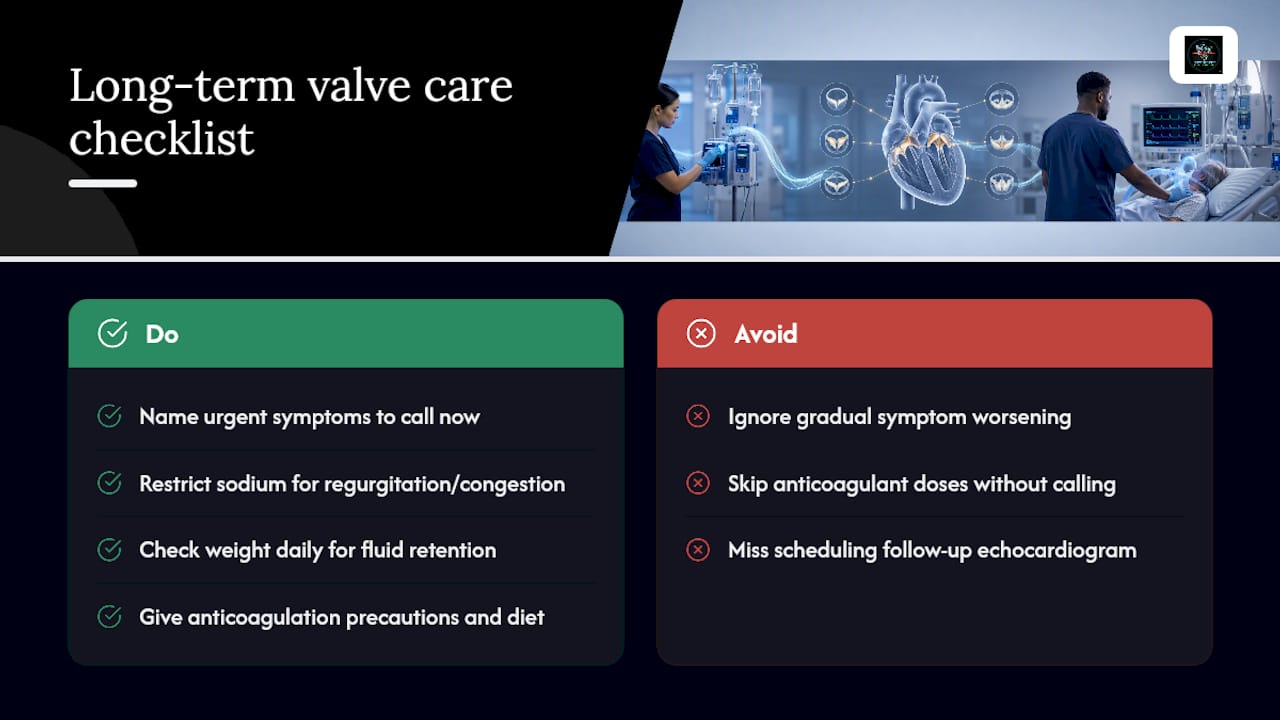
Teach patients to recognize the specific symptoms that mean “call your cardiologist now”: new or worsening shortness of breath, sudden weight gain from fluid retention, chest pain, fainting spells, or a racing or irregular heartbeat. The NHLBI notes that many valve disease symptoms develop slowly, which means patients often normalize gradual decline until they hit a crisis point. Your discharge teaching should explicitly name that risk.
Sodium restriction matters for anyone with regurgitant lesions or a history of pulmonary congestion, and daily weight checks catch fluid retention before it becomes dyspnea. Patients on anticoagulation after valve replacement need clear guidance on bleeding precautions, dietary interactions with warfarin if that’s their regimen, and the importance of never skipping doses without calling their prescriber first.
Endocarditis prophylaxis deserves a direct conversation for patients with prosthetic valves or a prior endocarditis history: they need to tell every dentist and proceduralist about their valve history, since certain procedures still warrant antibiotic prophylaxis under current guidance. Exercise isn’t universally restricted, but patients with severe stenosis need individualized activity guidance from their cardiologist, since exertion can unmask symptoms a resting exam misses. Reinforce follow-up echo scheduling too. Chronic valve disease is tracked over time, not diagnosed once and forgotten.
Nurse-to-Nurse: Common Pitfalls and Fast Mental Shortcuts
Don’t assume a chronic murmur means a stable patient. Watch for new vital sign changes. Use this shortcut: murmur timing tells you the lesion, the lesion tells you the hemodynamic shift, and that shift tells you your first action. Escalate fast on sudden hypotension, rising lactate despite support, or a new conduction block.
— Zero
Practice the Exact Reasoning AACN Tests
This question bank is built around the lesion-to-hemodynamics-to-action chain emphasized throughout this guide, aligning with the reasoning AACN tests on exam day. There are over 695 practice questions with detailed rationales, designed to help learners understand their mistakes rather than just mark answers incorrect.
Cardiovascular content, including valve disorders, gets its own dedicated question set alongside hemodynamic monitoring drills that train you to read PCWP and PA waveforms the way you’ll need to on exam day. If you’re closing in on a test date, the subscription page gives you full access through Premium Monthly at $14.99 per month, Yearly Access at $84.99 per year, or Lifetime Mastery for candidates who want one purchase and no renewal to track. Pick the plan that matches your timeline and start working through the cardiovascular set today.
Sources
These references anchor the clinical facts in this guide, and they’re worth revisiting as you build your own CCRN study notes.
- Heart Valve Diseases | NHLBI, NIH
- Aortic regurgitation — MedlinePlus
- Overview of cardiac valvular disorders | Merck Manual Professional
- CCRN exam handbook / test plan | AACN
This article is general information, not a substitute for advice from a qualified doctor. Consult a qualified healthcare professional about your own circumstances before acting on anything here.
FAQ
What Are the Four Types of Heart Valves?
The four heart valves are the tricuspid and mitral valves, which sit between the atria and ventricles, and the pulmonic and aortic valves, which control blood leaving the ventricles. The NHLBI identifies these as tricuspid, pulmonary, mitral (bicuspid), and aortic. Each can develop stenosis, regurgitation, prolapse, or, rarely, atresia.
What Are the Symptoms of Heart Valve Disease?
Common symptoms include fatigue, shortness of breath, chest pain, fainting spells, palpitations, and swelling in the legs or abdomen. According to the NHLBI, many people stay asymptomatic for years while the heart compensates, so new or worsening symptoms deserve prompt evaluation.
What Is the Life Expectancy for Someone With Mild Aortic Regurgitation?
Mild aortic regurgitation generally carries a favorable outlook when monitored with regular echocardiograms and managed appropriately, according to MedlinePlus. Outcomes depend on how the left ventricle tolerates the volume overload over time, which is why serial imaging matters more than a single snapshot.
What Is Heart Valve Disease?
Heart valve disease happens when one or more of the heart’s four valves can’t open or close properly, causing stenosis, regurgitation, prolapse, or atresia. The NHLBI frames it as a structural problem that disrupts normal blood flow through the heart, and CCRN candidates are expected to trace that disruption to its hemodynamic effects.
Does Zero Deficit™ Cover Valve Disorders for CCRN Prep?
Yes. Zero Deficit™ includes dedicated cardiovascular practice questions covering valve pathophysiology, hemodynamic interpretation, and management priorities. Full access, including cardiovascular modules, is available through the subscription plans.
Recommended
Take a Cardiovascular practice quiz
Test your mastery of hemodynamics, ACLS, MI, arrhythmias, and heart failure.
Get Your Free 20-Question CCRN Diagnostic
20 high-yield CCRN-style questions across every body system, with detailed rationales — emailed to you instantly.
No spam. One email + a couple of helpful follow-ups. Unsubscribe anytime.
Ready to pass your CCRN?
Join 12,000+ nurses using Zero Deficit — 695+ practice questions with rationales, AI-powered weak-area drilling, and spaced-repetition flashcards.
Start Free Trial →