The Role of Respiratory Assessment in Critical Care
TL;DR:
- Respiratory assessment systematically evaluates lung function to detect life-threatening conditions early.
- Performing a full five-step exam helps guide interventions and improves patient outcomes.
Respiratory assessment is the systematic process of evaluating a patient’s breathing and lung function to detect abnormalities that signal life-threatening conditions requiring immediate action. The role of respiratory assessment in critical care goes beyond counting breaths. It is the foundation for every ventilator decision, every diuresis call, and every code you prevent before it starts. Normal adult respiratory rate sits between 12–20 breaths per minute, with SpO2 between 94–98%. Any deviation from those baselines is your first clinical alarm. The SFMU and SRLF 2026 guidelines formalized this urgency with 20 consensus recommendations for managing acute respiratory failure, reinforcing that structured evaluation is not optional in the ICU.
What is the role of respiratory assessment in critical care?
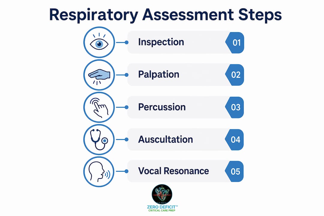
Respiratory assessment is a five-step clinical process: inspection, palpation, percussion, auscultation, and vocal resonance testing. Each step builds on the last, and skipping one creates a gap that can cost a patient their airway. You are not just collecting data. You are building a clinical picture that guides every intervention that follows.
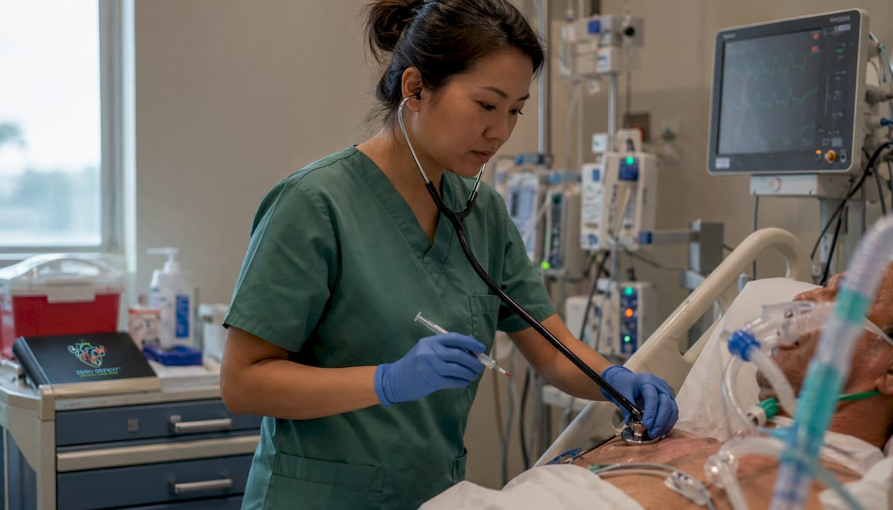
Inspection starts before you touch the patient. Count the respiratory rate for a full 60 seconds. Watch for accessory muscle use, nasal flaring, pursed-lip breathing, and asymmetrical chest rise. Cyanosis at the lips or fingertips tells you oxygenation has already failed at the tissue level.
Palpation confirms what your eyes suggest. Place your thumbs at the costal margin with fingers spread across the posterior chest. During deep inspiration, symmetrical expansion should move your thumbs at least 5 cm apart. Asymmetry points directly to unilateral pathology: pneumothorax, effusion, or lobar consolidation.
Percussion maps the density of underlying lung tissue. Dullness indicates fluid or consolidation. Hyper-resonance signals trapped air. Percussion technique requires strict side-to-side comparison without repeating the same spot, so subtle asymmetries do not get missed.
Auscultation demands patience. Listen through the full respiratory cycle anteriorly, posteriorly, and through the axillae. Vesicular sounds, bronchial sounds, crackles, and wheezes each carry a specific diagnostic message. Missing the axillae is one of the most common errors in clinical practice.
Vocal resonance adds a final layer. Ask the patient to say “ninety-nine” while you auscultate. Increased resonance indicates consolidation. Decreased resonance points to effusion or pneumothorax.
Pro Tip: Measure SpO2 on room air before applying supplemental oxygen whenever the clinical situation allows. That single number tells you where the patient’s baseline truly sits.
How does respiratory assessment aid in early detection of respiratory failure?
Early detection of respiratory failure depends on recognizing deviations from the patient’s own baseline, not just from population norms. Baseline SpO2 trends matter more than a single reading, particularly in COPD patients who may run saturation levels in the high 80s as their normal. Treating a COPD patient’s SpO2 of 88% as a crisis when it is their baseline leads to over-oxygenation and hypercapnic drive suppression.
Percussion and auscultation findings let you differentiate four major conditions at the bedside before imaging confirms anything.
| Condition | Percussion note | Breath sounds | Vocal resonance |
|---|---|---|---|
| Pneumonia | Dull | Bronchial, crackles | Increased |
| Pleural effusion | Stony dull | Absent | Decreased |
| Pneumothorax | Hyper-resonant | Absent | Decreased |
| COPD exacerbation | Hyper-resonant | Prolonged expiration, wheeze | Normal to decreased |
Distinct breath sounds and percussion notes map directly to these four conditions. That mapping is what separates a nurse who reacts to alarms from one who anticipates them.
The SFMU and SRLF 2026 framework reinforces integrating objective findings with patient history. A rising respiratory rate trend over two hours, combined with increasing accessory muscle use and a dropping SpO2, is acute respiratory failure in progress. You do not need a chest X-ray to act. You need a systematic clinical evaluation completed correctly.
- Watch for respiratory rate trends, not single readings
- Compare current findings to the patient’s documented baseline
- Integrate percussion, auscultation, and vital signs before calling the physician
- Document the full assessment so the next nurse has a real baseline to compare against
Pro Tip: In COPD patients, target SpO2 between 88–92% per current guidelines. Chasing 98% in this population can suppress their hypoxic drive and worsen CO2 retention.
What are common pitfalls in performing respiratory assessments?
The most damaging errors in respiratory assessment are not dramatic. They are quiet omissions that compound over time. Neglecting the posterior chest and axillae is the single most common failure in clinical practice. Lower lobe pneumonia and pleural effusions live in the bases. If you only auscultate the anterior chest, you will miss them every time.
Improper percussion technique is the second major pitfall. Repeating percussion on the same location instead of moving systematically side-to-side masks the asymmetry you are trying to find. The technique requires a consistent strike force and a methodical pattern from apex to base.
Incomplete auscultation cycles create false negatives. Some lung sounds only appear during inspiration, others only during expiration. Listening for half a cycle means half the data. Allow the patient to rest between auscultation cycles. Rapid shallow breathing from fatigue hides sounds that would otherwise be audible.
- Skipping the posterior chest exam
- Inconsistent percussion strike force
- Auscultating only the anterior chest
- Not allowing rest between breath cycles
- Overrelying on SpO2 without integrating the full clinical picture
Physical examination lacks standardized interpretation literature, which means expert mentorship fills a real gap. Working alongside experienced ICU nurses and respiratory therapists accelerates your ability to interpret findings accurately. Platforms like Mentoloop connect clinical learners with mentors who can provide real-time feedback on assessment technique, which is exactly the kind of guided practice that builds reliable clinical judgment.
Pro Tip: Always document what you heard, where you heard it, and during which phase of the respiratory cycle. Vague documentation like “breath sounds clear” is clinically useless and legally indefensible.
How can nurses master respiratory assessment for clinical and CCRN exam success?
Mastering respiratory evaluation techniques requires a structured framework you repeat until it becomes automatic. The OSCE-standard approach used in clinical education gives you that framework. It works in the ICU for the same reason it works in exams: it is systematic, reproducible, and complete.
- Start with the hands and face. Clubbing, cyanosis, and pursed-lip breathing give you a clinical context before you place your stethoscope.
- Assess respiratory rate and SpO2 on room air. Document both as your baseline before any intervention.
- Inspect the chest. Look for symmetry, accessory muscle use, and abnormal chest wall movement.
- Palpate for expansion. Use the thumb technique and measure for the 5 cm separation standard.
- Percuss systematically. Move side-to-side from apex to base, anterior and posterior.
- Auscultate completely. Cover anterior, posterior, and axillary regions through full respiratory cycles.
- Test vocal resonance. Ask the patient to speak and compare resonance bilaterally.
Practice with guided templates and checklists builds the muscle memory that holds under pressure. The CCRN respiratory study guide from Zerodeficitccrnprep maps this framework directly to AACN exam content, so you are not studying two separate things. You are reinforcing clinical skill and exam knowledge simultaneously.
Regular feedback from clinical educators accelerates skill development faster than solo practice. When you combine mentorship with targeted respiratory practice questions, you close the gap between knowing the steps and applying them correctly under pressure. That combination is what CCRN candidates who pass on their first attempt consistently report.
Key Takeaways
Systematic respiratory assessment is the most reliable early warning system in critical care, and every missed step is a missed diagnosis.
| Point | Details |
|---|---|
| Follow the five-step sequence | Inspection, palpation, percussion, auscultation, and vocal resonance must all be completed every time. |
| Use patient baseline, not population norms | SpO2 trends relative to the patient’s own baseline matter more than a single number, especially in COPD. |
| Auscultate posteriorly and through the axillae | Lower lobe pathology is missed when assessment stops at the anterior chest. |
| Integrate findings before acting | Percussion notes, breath sounds, and vital sign trends together build the clinical picture. |
| Combine clinical practice with targeted exam prep | Systematic assessment skill and CCRN exam success reinforce each other when studied together. |
Why respiratory assessment is the skill I never stop refining
I have watched nurses with five years of ICU experience miss a developing pleural effusion because they auscultated the anterior chest and called it done. I have also watched new graduates catch a tension pneumothorax before the attending because they followed the full sequence without cutting corners. The difference was not experience. It was discipline.
The uncomfortable truth about respiratory assessment is that most of us were taught it once and then left to practice it inconsistently. There is no standardized feedback loop in most ICUs. You do your assessment, document it, and move on. Nobody tells you that your percussion technique is too light or that you are not listening through full respiratory cycles. That gap between theory and practice is real, and it costs patients.
What I have found actually works is treating every assessment like an OSCE. Same sequence, same documentation, same standard every time. When you build that habit, you stop missing things. You also start noticing trends that others walk past. A respiratory rate that has climbed from 18 to 24 over four hours is a story. Most nurses miss it because they are looking at the current number, not the trend.
The 2026 SFMU and SRLF guidelines did not introduce anything revolutionary. They formalized what experienced critical care nurses already knew: structured assessment, applied consistently, saves lives. Your job is to internalize that structure until it runs on autopilot, and then use your clinical judgment on top of it.
— Zero
Sharpen your respiratory skills with Zerodeficitccrnprep
Respiratory assessment is one of the highest-yield topics on the AACN Adult CCRN exam. Getting it right in the ICU and getting it right on the exam require the same foundation: a systematic approach backed by current clinical guidelines.
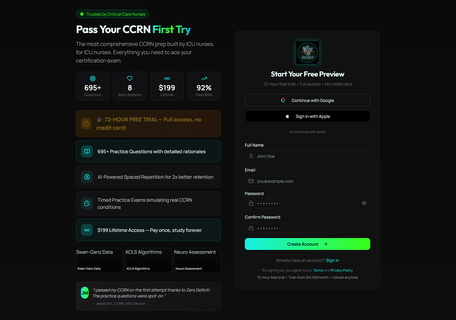
Zerodeficitccrnprep built the CCRN Respiratory Study Guide 2026 specifically for ICU nurses who need to master pulmonary concepts fast and retain them under exam pressure. The guide integrates the five-step assessment framework, ARDS management, ventilator weaning, and ABG interpretation into one focused resource. Pair it with the full CCRN study guide collection covering all eight body systems to build the complete clinical picture the CCRN exam tests. Every resource includes detailed rationales written by expert ICU nurses, so you understand the “why” behind every answer.
FAQ
What is the role of respiratory assessment in critical care?
Respiratory assessment is the systematic evaluation of breathing, lung sounds, chest expansion, and oxygenation to detect deterioration early and guide clinical intervention. It is the primary tool for identifying respiratory failure before it becomes a code.
What are the normal ranges for respiratory rate and SpO2?
Normal adult respiratory rate is 12–20 breaths per minute, and normal SpO2 is 94–98%. Deviations from these values, or from the patient’s personal baseline, signal possible respiratory compromise.
Why is auscultating the posterior chest and axillae important?
Posterior and axillary auscultation detects lower lobe pathology that anterior-only assessment misses entirely. Pleural effusions and basal pneumonia are the most common conditions caught only when the full chest is assessed.
How do percussion findings help differentiate respiratory conditions?
Dullness indicates consolidation or fluid, hyper-resonance indicates trapped air, and stony dullness points to pleural effusion. Combined with breath sounds and vocal resonance, percussion findings allow bedside differentiation of pneumonia, pneumothorax, and effusion.
How does respiratory assessment connect to CCRN exam preparation?
The CCRN exam tests clinical application of respiratory evaluation techniques, including ABG interpretation, ventilator management, and recognition of respiratory failure patterns. Mastering the systematic assessment process reinforces the exact knowledge the AACN exam blueprint tests in the pulmonary domain.
Recommended
Take a Respiratory practice quiz
Master ARDS, ventilation, ABGs, oxygenation, and pulmonary emergencies.
Get Your Free 20-Question CCRN Diagnostic
20 high-yield CCRN-style questions across every body system, with detailed rationales — emailed to you instantly.
No spam. One email + a couple of helpful follow-ups. Unsubscribe anytime.
Ready to pass your CCRN?
Join 12,000+ nurses using Zero Deficit — 695+ practice questions with rationales, AI-powered weak-area drilling, and spaced-repetition flashcards.
Start Free Trial →